 Auditory Scene
At a glance
Persistent inflammation of the middle ear and mastoid; the commonest form is chronic suppurative otitis media, with a permanent perforation and recurrent discharge.
Recurrent or persistent ear discharge (otorrhoea), conductive hearing loss and ear fullness; severe pain is usually absent.
Dizziness, facial palsy or severe headache are signs of complications and need emergency assessment.
Aural toilet and topical drops; surgery for perforation and cholesteatoma; hearing aids or bone conduction implants for persistent loss.
Medical disclaimer. This content is for informational purposes only and does not replace a physician’s examination, diagnosis or treatment; it should not be used as medical advice. For your complaints or personal situation, always consult an ear, nose and throat physician and an audiologist.
If you have recurrent or persistent discharge from your ear and your hearing is getting worse, see an ear, nose and throat doctor. If dizziness, facial weakness, severe headache or fever develop, seek emergency care without delay; these are rare but serious signs of complications.
Definition and epidemiology
Chronic otitis media (COM) is persistent inflammation of the middle ear and mastoid. Its commonest form is chronic suppurative otitis media (CSOM), with a permanent hole in the eardrum (tympanic membrane perforation) and intermittent discharge. Cholesteatoma, an accumulation of keratinising epithelium in the middle ear, is considered under this heading as the squamous form of COM.
According to the World Health Organization, CSOM is a major cause of preventable childhood hearing loss, particularly in low-income regions; a community prevalence above 4% is regarded as a public-health problem requiring urgent action (WHO, 2004).
The disease usually becomes chronic through a combination of Eustachian tube dysfunction, recurrent infection and persistent changes in the middle-ear mucosa (Verhoeff et al., 2006).
Symptoms and signs
The most typical symptoms are recurrent or persistent ear discharge (otorrhoea), conductive hearing loss and occasional ear fullness. Unlike acute otitis media, severe pain is usually absent.
The painless course can be misleading: ossicular damage or cholesteatoma may progress unnoticed. Long-standing discharge and hearing loss should therefore always be assessed.
Dizziness, facial palsy and severe headache are rare but serious signs of complications and require emergency assessment.
Causes and risk factors
Eustachian tube dysfunction and recurrent middle-ear infection lie in the background. Previous episodes of acute otitis media, a perforation that fails to heal and chronic inflammation of the middle-ear mucosa keep the process going (Mittal et al., 2015).
Frequent upper respiratory infections, tobacco smoke, crowded living conditions, poor access to healthcare and craniofacial anomalies such as cleft palate increase the risk.
Cholesteatoma may be congenital, but it usually develops as an acquired process on a background of Eustachian tube dysfunction and a retraction pocket; because it is progressive and can erode surrounding bone, it is of particular importance (Kuo et al., 2015).
Audiological and clinical assessment
Assessment is based on otoscopic and microscopic examination, which shows the perforation, discharge and signs of cholesteatoma directly. High-resolution CT of the temporal bone is used when cholesteatoma or a complication is suspected, and diffusion-weighted MRI when looking for residual or recurrent cholesteatoma after surgery.
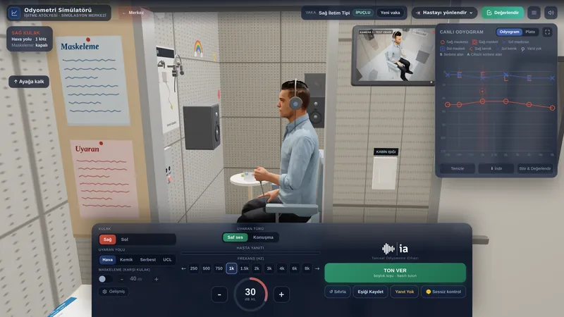
- Pure-tone audiometry: establishes the degree of conductive or mixed loss and the air-bone gap; bone-conduction thresholds show inner-ear involvement.
- Tympanometry: with a perforation, a flat trace with a high ear-canal volume; with an intact drum, information on middle-ear pressure and mobility.
- Speech audiometry: the speech discrimination score helps to assess inner-ear function and rehabilitation options.
- Masking: often needed for accurate bone-conduction thresholds in unilateral or asymmetric conductive loss.
Audiological findings guide both surgical planning and candidacy for hearing aids or bone conduction implants.
Treatment and audiological rehabilitation
Active discharge is treated first with aural toilet and appropriate topical antibiotic drops (Mittal et al., 2015); non-ototoxic drops are preferred when the drum is perforated. A persistent perforation, ossicular damage or cholesteatoma brings surgery (tympanoplasty, with mastoidectomy where needed) into consideration; the decision rests with the surgeon.
In cholesteatoma the main aim is to remove the disease completely and prevent complications; restoring hearing is secondary and is often addressed at a second stage.
Hearing aids can be used by patients with persistent hearing loss despite surgery or who are not suitable for surgery. If ear-canal discharge or ear anatomy makes a conventional aid unsuitable, bone conduction implants are considered.
Impact on quality of life and advice
Recurrent discharge and hearing loss can affect school and working life, social communication and self-confidence. In children, untreated hearing loss can hold back language development.
If your eardrum is perforated, keep water out of the ear, seek help early when discharge flares up and keep your follow-up appointments. Follow-up after surgery matters for detecting recurrence early.
If you used this review, you can cite it as follows (APA 7):
Auditory Scene. (2026). Chronic Otitis Media. Hearing & Balance Health Guide. https://auditoryscene.com/guncel-haberler/categories/isitme-sagligi-rehberi/kronik-otitis-media/Permanent link: auditoryscene.com/guncel-haberler/categories/isitme-sagligi-rehberi/kronik-otitis-media/ · Last reviewed: October 2026 · License: CC BY-NC-ND 4.0
References
- Kuo, C. L., Shiao, A. S., Yung, M., et al. (2015). Updates and knowledge gaps in cholesteatoma research. BioMed Research International, 2015, 854024.
- Mittal, R., Lisi, C. V., Gerring, R., et al. (2015). Current concepts in the pathogenesis and treatment of chronic suppurative otitis media. Journal of Medical Microbiology, 64(10), 1103-1116.
- Verhoeff, M., van der Veen, E. L., Rovers, M. M., et al. (2006). Chronic suppurative otitis media: A review. International Journal of Pediatric Otorhinolaryngology, 70(1), 1-12.
- World Health Organization. (2004). Chronic suppurative otitis media: Burden of illness and management options. World Health Organization.
Frequently asked questions
I have constant discharge from my ear; is it dangerous?
Long-standing ear discharge can be a sign of chronic middle-ear inflammation and should be assessed by an ENT doctor. Most cases can be controlled with appropriate treatment; if neglected, hearing loss and, rarely, serious complications can develop.
Will the hole in my eardrum close on its own?
Small holes may sometimes heal on their own, but large, chronic holes usually require surgical repair (tympanoplasty). The state of the hole and its effect on hearing become clear with your physician’s examination.
Is cholesteatoma cancer?
No, cholesteatoma is not cancer. However, it is a skin accumulation that can grow in the middle ear and damage the surrounding bone. Because of its progressive nature it should not be neglected and usually needs to be removed surgically.
Will surgery restore my hearing?
The main aim of some operations is to remove the disease and make the ear safe; improving hearing depends on the state of the ossicles and is sometimes done in a second stage. It is important to clarify your expectations with your physician before surgery.
What happens if water gets into my ear?
In people with a drum perforation, water reaching the middle ear can trigger infection. Using the protective methods your physician recommends during bathing and swimming reduces discharge episodes.
Scales that can be used to monitor the impact of hearing loss on daily life and, in children, on development: