Auditory Scene
At a glance
Hidden hearing loss is a reduction in the information passed from the inner ear to the auditory nerve despite a normal audiogram; its best-known cause is loss of inner hair cell synapses (cochlear synaptopathy).
The main complaint is difficulty following speech in noise; tinnitus and heightened sensitivity to sound may accompany it.
It is not an emergency, but sudden hearing loss, persistent tinnitus or one-sided symptoms need medical assessment.
No clinical test can confirm the diagnosis; thorough audiological assessment, counselling and noise protection are the core of management.
Medical disclaimer. This content is for informational purposes only and does not replace a physician’s examination, diagnosis or treatment; it should not be used as medical advice. For your complaints or personal situation, always consult an ear, nose and throat physician and an audiologist.
If your audiogram is normal but you struggle to follow speech in noisy places, or you have tinnitus or heightened sensitivity to sound, tell your audiologist. If muffled hearing and ringing after noise exposure do not settle within a few days, if your symptoms are clearly worse in one ear, or if your hearing drops suddenly, see an ear, nose and throat doctor without delay.
Definition and epidemiology
Hidden hearing loss describes conditions in which pure-tone thresholds are normal but the information passed from the inner ear to the auditory nerve is reduced or degraded. The term became widely used after a small ABR wave I, generated by the auditory nerve, was found in people with tinnitus and normal audiograms (Schaette and McAlpine, 2011). The loss is ‘hidden’ because the standard audiogram measures only threshold; it says nothing about how faithfully suprathreshold sounds are coded in the nerve.
Cochlear synaptopathy is the loss of the ribbon synapses between inner hair cells and auditory nerve fibres while the hair cells themselves survive. Although hidden hearing loss is often used as a synonym, it is a broader concept: disorders of auditory nerve myelin, peripheral neuropathies and some ototoxic effects can produce a similar picture (Kohrman et al., 2020). Its prevalence in humans is unknown, because synaptopathy cannot be demonstrated directly in a living person; conclusive evidence can come only from temporal bone examination (Bramhall et al., 2019).
Symptoms and signs
The commonest complaint is from someone who manages well in quiet but struggles to follow conversation in crowded, noisy or reverberant settings. People may describe fatigue after long listening, or say they can ‘hear but not make out’ speech. In university students with normal audiograms, those at high risk from noise exposure scored lower on word recognition in noise and with time-compressed or reverberant speech, and reported more reactions to sound (Liberman et al., 2016).
Tinnitus may be present. In people with tinnitus and normal audiograms, ABR wave I amplitude was smaller than in controls while wave V was normal, interpreted as reduced nerve input being offset by increased gain in the brainstem (Schaette and McAlpine, 2011). For heightened sensitivity to sound, see the entry on decreased sound tolerance. None of these complaints is specific to synaptopathy; attention, memory and language skills also affect listening in noise (Bramhall et al., 2019).
Causes and risk factors
The key animal finding comes from the mouse study by Kujawa and Liberman (2009). In mice exposed for 2 hours to 8-16 kHz noise at 100 dB SPL, thresholds rose by about 40 dB at 24 hours and returned to normal by 2 weeks. There was no hair cell loss and DPOAEs recovered, yet about half of the inner hair cell synapses in the most affected region were lost and had not returned 8 weeks later. ABR wave I amplitude stayed reduced, and spiral ganglion cells fell by about 50% over 2 years. This challenged the assumption that a ‘temporary’ threshold shift is harmless.
Animal studies suggest that noise mainly affects high-threshold nerve fibres (Plack et al., 2014). These fibres do not set the threshold in quiet but help code loud and noisy sounds, so thresholds can remain normal while hearing in noise suffers. Ageing also causes synaptopathy: in a study of 20 temporal bones from people aged 0-86 years, inner hair cell loss rarely exceeded 15%, whereas 7 of the 11 people over 60 had lost more than 60% of peripheral nerve axons (Wu et al., 2019). Myelin is another target: transient loss of Schwann cells in mice caused a permanent reduction in wave I with no change in synapse numbers (Wan and Corfas, 2017).
Human evidence for noise-induced synaptopathy is inconsistent. According to the review by Bramhall et al. (2019), some studies have linked noise history with a smaller wave I, while most studies in people with normal audiograms have not. Possible reasons include lower human susceptibility compared with rodents, large individual variability in evoked potentials, the unreliability of questionnaire-based noise histories and subclinical outer hair cell damage.
Audiological and clinical assessment
No clinical test can confirm hidden hearing loss; without temporal bone histology no measure can separate synaptopathy from other pathology (Bramhall et al., 2019). In a person with symptoms and a normal audiogram, the audiologist aims to exclude organic causes, measure suprathreshold hearing and document the complaint objectively.
- Pure-tone and extended high-frequency audiometry: thresholds from 250 to 8000 Hz are normal by definition. Testing above 8 kHz can reveal early damage that the standard audiogram misses: students at high risk from noise had significantly raised thresholds at 10-16 kHz (Liberman et al., 2016). Bramhall et al. (2019) recommend testing up to 12-16 kHz, although such a finding is not specific to synaptopathy.
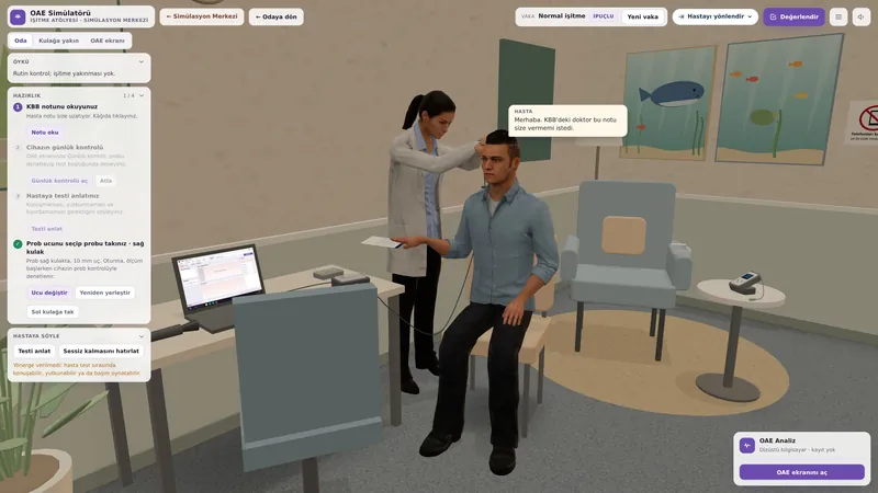
- Tympanometry and DPOAEs: showing normal middle ear and outer hair cell function supports a neural origin for the complaint. DPOAEs should be interpreted beyond a simple pass or fail, including at low stimulus levels (Bramhall et al., 2019).
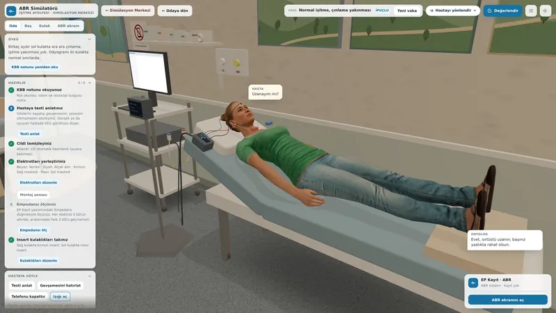
- ABR wave I amplitude: the most widely used measure. Recording with high-level stimuli (90-100 dB peSPL) and enough averages is recommended (Bramhall et al., 2019). Individual variability is large and outer hair cell damage affects the result, so normal limits are hard to set for a single person. The wave V to wave I ratio is being studied as a marker of increased central gain (Schaette and McAlpine, 2011).
- Electrocochleography (ECochG): the ratio of the summating potential to the action potential (SP/AP) differed in a high-risk group and has been proposed as a marker of selective neural loss (Liberman et al., 2016).
- Middle ear muscle reflex (MEMR): because the acoustic reflex pathway relies on high-threshold fibres, it is being studied as a measure sensitive to synaptopathy; weaker reflexes have been reported in people with tinnitus, but audiometric differences between groups may have contributed (Bramhall et al., 2019).
- Speech-in-noise tests: these document the complaint objectively and are valuable for follow-up. In Turkish, the matrix sentence test, HINT-TR and QuickSIN-TR are available. Because results also depend on attention, memory and language, they are not markers of synaptopathy on their own.
| Measure | Possible finding | Limitation |
|---|---|---|
| ABR wave I amplitude | Small wave I at high levels | Large individual variability |
| Wave V/I ratio | Increased central gain | Indirect marker |
| SP/AP ratio (ECochG) | Altered ratio | Not widely used clinically |
| MEMR | Weak reflex | Affected by audiometric differences |
| Extended high frequencies | Raised thresholds at 10-16 kHz | Not specific to synaptopathy |
| Speech in noise | Low scores | Affected by attention and memory |
| DPOAEs | Normal | To exclude outer hair cell damage |
The differential diagnosis includes auditory processing disorder, ANSD and early noise-induced hearing loss or presbycusis. In ANSD, OAEs are present while the ABR is absent or grossly abnormal; in hidden hearing loss the ABR waves are present and only wave I may be small. In auditory processing disorder the problem lies in the auditory pathways above the brainstem. If symptoms are clearly worse in one ear, medical assessment is needed to exclude retrocochlear disease.
Treatment and audiological rehabilitation
There is no proven treatment. Hearing aids and cochlear implants mainly address threshold loss and do not restore lost synapses (Kohrman et al., 2020). In animal studies, molecules such as neurotrophin-3 given after noise exposure have promoted synapse regeneration, but these approaches are not yet in clinical use in humans (Kohrman et al., 2020). Decisions on medication and experimental treatment rest with the doctor.
Audiological care begins by taking the complaint seriously. Someone told that their hearing is normal may conclude their difficulty is not real, yet difficulty hearing in noise is a measurable and explainable problem. Counselling covers communication strategies (facing the speaker, sitting away from noise sources, choosing well-lit settings), breaks to reduce listening effort and, where needed, assistive listening devices such as a remote microphone system. Evidence for these devices in hidden hearing loss is limited. If tinnitus or sound sensitivity is prominent, the principles of tinnitus management apply.
Prevention is the main approach. Animal findings show that noise can leave permanent neural damage even when thresholds recover (Kujawa and Liberman, 2009). Hearing protection in noisy workplaces, earplugs at concerts and venues, limiting volume and listening time on personal music players, and audiological monitoring for people on ototoxic drugs are recommended. Anyone who has had muffled hearing or ringing after noise should take more care at the next exposure, even if their thresholds recovered.
Impact on quality of life and advice
For someone with suspected hidden hearing loss, the hardest situations are busy restaurants, meetings, classrooms and noisy workplaces. A person who communicates without difficulty in quiet but struggles in these settings may be seen by others as inattentive or uninterested. Prolonged listening can lead to fatigue, withdrawal from social situations and reduced productivity at work.
That the concept is debated does not mean the complaint is not real. Although the diagnosis and frequency of synaptopathy in humans remain uncertain, difficulty hearing in noise can be measured, followed over time and eased with communication strategies. For someone whose symptoms persist despite a normal audiogram, repeating the assessment every few years allows any threshold loss to be picked up early.
In noisy places, face the person speaking, choose a quiet corner where possible and hold important conversations away from noise. Wear hearing protection in noisy jobs and at concerts, and keep the volume low and listening time limited on personal music players. If your symptoms worsen or tinnitus starts, see your audiologist.
If you used this review, you can cite it as follows (APA 7):
Auditory Scene. (2026). Hidden Hearing Loss (Cochlear Synaptopathy). Hearing & Balance Health Guide. https://auditoryscene.com/guncel-haberler/categories/isitme-sagligi-rehberi/gizli-isitme-kaybi/Permanent link: auditoryscene.com/guncel-haberler/categories/isitme-sagligi-rehberi/gizli-isitme-kaybi/ · Last reviewed: October 2026 · License: CC BY-NC-ND 4.0
References
- Bramhall, N., Beach, E. F., Epp, B., et al. (2019). The search for noise-induced cochlear synaptopathy in humans: Mission impossible? Hearing Research, 377, 88-103.
- Kohrman, D. C., Wan, G., Cassinotti, L., et al. (2020). Hidden hearing loss: A disorder with multiple etiologies and mechanisms. Cold Spring Harbor Perspectives in Medicine, 10(1), a035493.
- Kujawa, S. G., & Liberman, M. C. (2009). Adding insult to injury: Cochlear nerve degeneration after “temporary” noise-induced hearing loss. The Journal of Neuroscience, 29(45), 14077-14085.
- Liberman, M. C., Epstein, M. J., Cleveland, S. S., et al. (2016). Toward a differential diagnosis of hidden hearing loss in humans. PLoS ONE, 11(9), e0162726.
- Plack, C. J., Barker, D., & Prendergast, G. (2014). Perceptual consequences of “hidden” hearing loss. Trends in Hearing, 18, 2331216514550621.
- Schaette, R., & McAlpine, D. (2011). Tinnitus with a normal audiogram: Physiological evidence for hidden hearing loss and computational model. The Journal of Neuroscience, 31(38), 13452-13457.
- Wan, G., & Corfas, G. (2017). Transient auditory nerve demyelination as a new mechanism for hidden hearing loss. Nature Communications, 8, 14487.
- Wu, P. Z., Liberman, L. D., Bennett, K., et al. (2019). Primary neural degeneration in the human cochlea: Evidence for hidden hearing loss in the aging ear. Neuroscience, 407, 8-20.
Frequently asked questions
My audiogram is normal but I cannot follow speech in noise. Is this hidden hearing loss?
It may be, but it is not the only explanation. Difficulty in noise can also relate to attention, memory, language skills, auditory processing problems or losses at high frequencies that the standard audiogram does not measure. Your audiologist can assess the complaint in detail with extended high-frequency audiometry, OAEs, ABR and speech-in-noise tests.
Can hidden hearing loss be diagnosed with certainty?
Not at present. Synapse loss can only be shown directly by microscopic examination of the temporal bone. Measures such as ABR wave I amplitude, the middle ear muscle reflex and electrocochleography are being studied, but none is reliable enough to make the diagnosis in an individual.
The ringing and muffled hearing after a concert went away. Could there be lasting damage?
Animal studies have shown that noise can leave permanent damage to the synapses of the auditory nerve even when thresholds return fully to normal. How far this applies to humans is not certain. Treat the experience as a warning and wear earplugs at future concerts. If the symptoms do not settle within a few days, see a doctor.
Is there a treatment? Do hearing aids help?
There is no proven treatment. Because hearing aids target threshold loss, they are not usually recommended for someone with a normal audiogram. Drugs aimed at regrowing synapses look promising in animals but are not in human use. Communication strategies and, for some people, assistive devices such as a remote microphone system can help.
Is hidden hearing loss the same as auditory processing disorder?
No. In hidden hearing loss the problem lies in the connections between the inner ear and the auditory nerve; in auditory processing disorder it lies in the brainstem and higher auditory pathways. Both can occur with a normal audiogram and difficulty in noise; the history and test battery tell them apart.
How can I protect myself?
Protecting your ears from noise is the most effective step. Use hearing protection in noisy workplaces and earplugs at concerts and venues, and keep the volume low and listening time limited with headphones. If you take ototoxic medication, ask for your hearing to be monitored.
Turkish tests that can be used to measure and monitor difficulty hearing speech in noise objectively: