 Auditory Scene
At a glance
Fluid collects in the middle ear without signs of acute infection; Eustachian tube dysfunction lies behind it.
Muffled hearing, turning up the television and not responding when called stand out; ear fullness and sometimes balance problems may accompany them.
Not an emergency; assessment is needed if the fluid lasts longer than 3 months or affects language development.
A child without risk factors is observed for 3 months; if the fluid persists and hearing is affected, ventilation tubes (grommets) are inserted, with adenoidectomy in selected children.
Medical disclaimer. This content is for informational purposes only and does not replace a physician’s examination, diagnosis or treatment; it should not be used as medical advice. For your complaints or personal situation, always consult an ear, nose and throat physician and an audiologist.
If your child hears muffled sounds, turns up the television, does not respond when called, or is inattentive in class, consult an ear, nose and throat physician and an audiologist. Children with Down syndrome, cleft palate, autism spectrum disorder or existing hearing loss need closer follow-up.
Definition and epidemiology
Otitis media with effusion (OME), often called glue ear, is the collection of fluid in the middle-ear cavity without the typical signs of acute infection. It stems from impaired ventilation and drainage by the Eustachian tube; the resulting negative pressure favours fluid build-up. Fluid can also persist after an episode of acute otitis media.
About 90% of children have OME at least once before school age. Most episodes resolve spontaneously within 3 months, but 30-40% of children have repeated episodes and 5-10% of episodes last a year or longer (Rosenfeld et al., 2016). In adults, unilateral persistent OME calls for separate examination of the nasopharynx.
Fluid in the middle ear hinders the transmission of sound to the inner ear and usually causes a mild-to-moderate conductive hearing loss.
Symptoms and signs
OME is often silent. Severe pain and fever are not expected; instead, hearing-related signs stand out, such as hearing sounds as muffled, turning up the television, not responding when called, or inattention in class.
Older children and adults may describe ear fullness, pressure or a feeling of water in the ear. Parents report balance symptoms in about 15% of children with OME, and clumsiness may also be seen (Rosenfeld et al., 2016).
Because the signs are subtle, diagnosis can be delayed; this can affect speech and language development, especially in young children.
Causes and risk factors
Eustachian tube dysfunction underlies it. Upper respiratory infections, allergy, enlarged adenoids and previous episodes of acute otitis media are the main triggers.
Nursery attendance, tobacco smoke, bottle-feeding while lying down, crowded living and the winter rise in infections increase the risk.
Children with permanent hearing loss independent of OME, speech and language delay, autism spectrum disorder, syndromes such as Down syndrome or craniofacial disorders, cleft palate, uncorrectable visual impairment or developmental delay are considered at risk; they are more vulnerable to the developmental effects of OME and are followed more closely (Rosenfeld et al., 2016).
Audiological and clinical assessment
OME is diagnosed with pneumatic otoscopy; because it shows eardrum mobility, it is more reliable than visual inspection alone. Tympanometry is used when the diagnosis is uncertain (Rosenfeld et al., 2016).
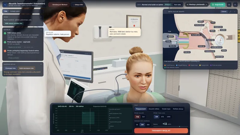
- Tympanometry: shows middle-ear fluid objectively; OME typically gives a flat (Type B) trace with a normal ear-canal volume.
- Probe-tone choice: a 1000 Hz probe tone is recommended in infants under 6 months; the standard 226 Hz can be misleading (Rosenfeld et al., 2016).
- Audiometry: an age-appropriate hearing test is done if the fluid persists for 3 months or longer, in at-risk children, and whenever hearing or language development is in doubt (Rosenfeld et al., 2022).
- OAEs and acoustic reflexes: usually absent while fluid is present; OME is a common cause of a ‘refer’ result in newborn screening.
- Acoustic reflectometry: may be supportive in selected situations.
Routine screening for OME is not recommended in children who are not at risk and have no symptoms; assessment is prompted by symptoms or risk factors (Rosenfeld et al., 2016).
| Feature | Acute (AOM) | With effusion (OME) | Chronic (COM) |
|---|---|---|---|
| Pain/fever | Prominent | None/mild | Usually none |
| Middle-ear fluid | Inflamed (purulent) | No signs of acute inflammation | Recurrent discharge |
| Eardrum | Bulging, red | Retracted/dull | Permanent perforation possible |
| Hearing | Transient decline | Mild-moderate conductive | Conductive/mixed loss |
| First approach | Pain relief, antibiotic if needed | Watchful waiting (3 months) | Aural toilet, drops, surgery if needed |
Treatment and audiological rehabilitation
Guidelines advise against antibiotics, intranasal or systemic steroids, antihistamines and decongestants in OME, as no lasting benefit has been shown (Rosenfeld et al., 2016; Simon et al., 2018).
Children without risk factors are observed for 3 months. If bilateral OME lasts 3 months or longer and hearing difficulty is documented, bilateral ventilation tubes are recommended; children with chronic OME who do not have tubes are re-examined every 3-6 months until the fluid clears or significant hearing loss or structural changes of the eardrum are found (Rosenfeld et al., 2022). Under 4 years of age, adenoidectomy is not performed without a separate indication; from 4 years, tubes, adenoidectomy or both are options (Rosenfeld et al., 2016). The effect of adenoidectomy on hearing is uncertain; it may slightly reduce persistence of the effusion, but the certainty of the evidence is very low (MacKeith et al., 2023).
When surgery is unsuitable or not wanted, hearing aids, a remote microphone system in class and acoustic adjustments are considered.
Impact on quality of life and advice
Fluctuating hearing, with the child seeming to hear at some times and not at others, can worry the family and cause attention and communication difficulties for the child. Recognising it early protects language development.
Keeping the home smoke-free, speaking to the child face to face and clearly, and seating them near the front in class increase auditory input. Do not skip follow-up appointments.
If you used this review, you can cite it as follows (APA 7):
Auditory Scene. (2026). Otitis Media with Effusion (Serous Otitis). Hearing & Balance Health Guide. https://auditoryscene.com/guncel-haberler/categories/isitme-sagligi-rehberi/efuzyonlu-otitis-media/Permanent link: auditoryscene.com/guncel-haberler/categories/isitme-sagligi-rehberi/efuzyonlu-otitis-media/ · Last reviewed: October 2026 · License: CC BY-NC-ND 4.0
References
- MacKeith, S., Mulvaney, C. A., Galbraith, K., et al. (2023). Adenoidectomy for otitis media with effusion (OME) in children. Cochrane Database of Systematic Reviews, 2023(10), CD015252.
- Rosenfeld, R. M., Shin, J. J., Schwartz, S. R., et al. (2016). Clinical practice guideline: Otitis media with effusion (update). Otolaryngology-Head and Neck Surgery, 154(1_suppl), S1-S41.
- Rosenfeld, R. M., Tunkel, D. E., Schwartz, S. R., et al. (2022). Clinical practice guideline: Tympanostomy tubes in children (update). Otolaryngology-Head and Neck Surgery, 166(1_suppl), S1-S55.
- Simon, F., Haggard, M., Rosenfeld, R. M., et al. (2018). International consensus (ICON) on management of otitis media with effusion in children. European Annals of Otorhinolaryngology, Head and Neck Diseases, 135(1S), S33-S39.
Frequently asked questions
Why isn’t my child given antibiotics?
The fluid in otitis media with effusion is mostly not a microbial infection; antibiotics therefore provide no lasting benefit in clearing the fluid and carry side-effect and resistance risks if used unnecessarily. That is why current guidelines recommend waiting and follow-up.
Will waiting 3 months set my child’s speech back?
In children without developmental risk factors, 3 months of observation is considered safe, because most cases resolve in this time. During this period it is important to talk face to face with the child a lot and support their hearing; if symptoms persist, a hearing test is done.
How do grommets affect speech development?
Grommets ventilate the middle ear, and in most children hearing improves while the tube is in place. This helps the child hear speech more clearly during a period of rapid language development; evidence for a long-term effect on language, however, is limited.
Can my child swim after grommets are fitted?
Yes. The current guideline does not recommend avoiding swimming or routine precautions such as earplugs or headbands for children with tubes (Rosenfeld et al., 2022). If the ear starts to discharge, contact your doctor.
Will the fluid build up again?
Yes, effusion can recur, especially in young children and those with frequent upper respiratory infections. Regular check-ups are therefore recommended while the tube is in place and after it comes out.
Scales that can be used to monitor Eustachian tube function and, in children, the impact of hearing loss: