 Auditory Scene
At a glance
Remodelling of the bone around the stapes; the stapes becomes fixed and hearing loss develops.
Slowly progressive hearing loss and tinnitus are typical; some patients hear speech relatively better in noisy places (paracusis Willisii).
Not an emergency, but because it is progressive it needs regular audiological follow-up.
Stapes surgery (stapedotomy or stapedectomy) or a hearing aid; the choice is made together with the patient.
Medical disclaimer. This content is for informational purposes only and does not replace a physician’s examination, diagnosis or treatment; it should not be used as medical advice. For your complaints or personal situation, always consult an ear, nose and throat physician and an audiologist.
If you notice slowly worsening hearing in one ear, a family history of hearing loss, tinnitus or relatively better hearing in noisy places, see an ear, nose and throat doctor and an audiologist. If there is a sudden change in hearing, dizziness or rapid worsening in one ear, seek assessment promptly.
Definition and epidemiology
Otosclerosis is a localised bone-remodelling disease specific to the otic capsule surrounding the inner ear. Normal bone is replaced by irregular, vascular, spongy bone. The process most often involves the fissula ante fenestram, just in front of the oval window (Quesnel et al., 2018).
When this change fixes the stapes footplate, sound vibrations are not transmitted adequately to the inner ear and a conductive hearing loss results. If the focus extends into the cochlea, a sensorineural component is added and a mixed loss can develop.
Clinical otosclerosis is more common in women and usually begins in early adulthood; in a five-centre British cohort, 65% of patients were women (Crompton et al., 2019).
Symptoms and signs
The commonest symptom is a slowly progressive hearing loss that often starts in one ear and may become bilateral over time. The loss usually affects the low frequencies first.
Many patients also have tinnitus. Some report understanding speech relatively better in noisy places; this finding, known as paracusis Willisii, is seen in conductive losses.
Otosclerosis begins insidiously, without signs of infection such as pain, discharge or redness. The eardrum usually looks normal; a conductive loss in a normal-looking ear points the clinician towards otosclerosis.
Causes and risk factors
Otosclerosis is multifactorial; genetic predisposition is thought to act together with environmental and hormonal factors. Both familial and sporadic forms occur.
In the same cohort, 40% of patients reported a family history, and familial cases had an earlier onset (Crompton et al., 2019). Gene variants related to bone remodelling and a possible link with measles virus have been studied, but the cause has not been fully explained (Chole and McKenna, 2001).
Women who link their hearing loss to pregnancy have been reported to have an earlier onset, suggesting that pregnancy may accelerate progression in some women (Crompton et al., 2019).
Audiological and clinical assessment
Diagnosis rests on ENT examination together with the audiological findings. A conductive loss despite a normal-looking eardrum is typical.
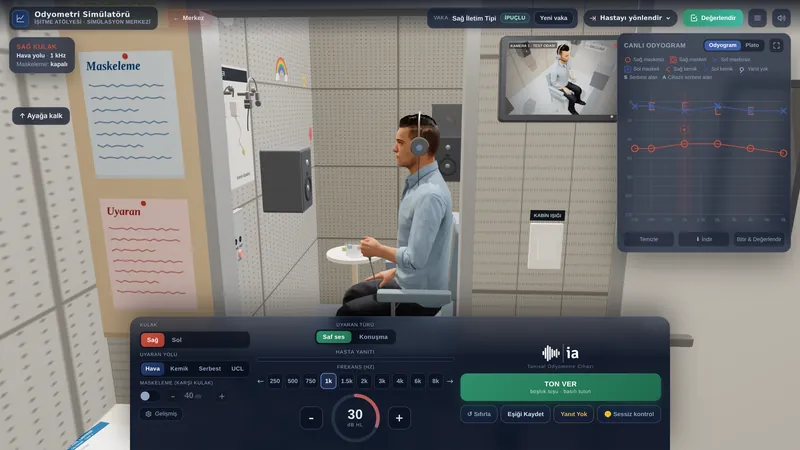
- Pure-tone audiometry: a conductive (in advanced stages mixed) loss with an air-bone gap.
- Carhart notch: a dip in bone-conduction thresholds around 2 kHz (Carhart, 1950). It is a mechanical effect of stapes fixation, often resolves after successful surgery and is not diagnostic on its own.
- Tympanometry: usually normal (Type A) or low-peaked (Type As), reflecting middle-ear stiffness.
- Acoustic reflexes: weakened or lost early; absent once fixation is established.
Wideband tympanometry (WBT) can show middle-ear stiffening in more detail. High-resolution CT is used to assess the extent of the foci when surgery is planned (Quesnel et al., 2018). The differential diagnosis includes tympanosclerosis and superior semicircular canal dehiscence (SSCD); SSCD can also give an air-bone gap with a normal drum, but the acoustic reflexes are preserved.
| Feature | Otosclerosis | Tympanosclerosis | SSCD |
|---|---|---|---|
| Main involvement | Stapes and oval window | Calcification of drum and ossicles | Superior semicircular canal (third window) |
| Eardrum | Looks normal | White plaques may be seen | Looks normal |
| Acoustic reflex | Lost early | May be lost | Preserved |
| OAE | May be affected | May be affected | Preserved |
| Distinguishing clue | Carhart notch, family history | History of infection or tubes | Autophony, sound-induced vertigo, low VEMP thresholds |
Treatment and audiological rehabilitation
In surgery, part of the fixed stapes is removed and replaced with a small prosthesis (stapedotomy or stapedectomy). In experienced hands the success rate for closing the air-bone gap is high; permanent sensorineural hearing loss is a rare but important risk. The decision is made by the surgeon together with the patient.
Hearing aids are effective for patients who do not want, or are not suitable for, surgery. Because the inner ear is intact in conductive loss, speech discrimination is usually good.
In selected cases where the disease has extensively involved the cochlea, a far-advanced mixed loss has developed and hearing aids no longer give enough benefit, a cochlear implant may be considered. Stapes surgery is increasingly complemented, or replaced, by advanced hearing aids and cochlear implants (Batson and Rizzolo, 2017).
Impact on quality of life and advice
Hearing loss can lead to listening fatigue, social withdrawal and anxiety. Treatment success should be judged not only by the audiogram but also by how comfortably the person communicates in daily life.
Because the disease is progressive, regular audiological check-ups are important. Family members who understand the condition, and strategies such as speaking face to face and clearly in noisy settings, ease the burden on the patient.
If you used this review, you can cite it as follows (APA 7):
Auditory Scene. (2026). Otosclerosis. Hearing & Balance Health Guide. https://auditoryscene.com/guncel-haberler/categories/isitme-sagligi-rehberi/otoskleroz/Permanent link: auditoryscene.com/guncel-haberler/categories/isitme-sagligi-rehberi/otoskleroz/ · Last reviewed: October 2026 · License: CC BY-NC-ND 4.0
References
- Batson, L., & Rizzolo, D. (2017). Otosclerosis: An update on diagnosis and treatment. JAAPA, 30(2), 17-22.
- Carhart, R. (1950). Clinical application of bone conduction audiometry. Archives of Otolaryngology, 51(6), 798-808.
- Chole, R. A., & McKenna, M. (2001). Basic science review: Pathophysiology of otosclerosis. Otology & Neurotology, 22(2), 249-257.
- Crompton, M., Cadge, B. A., Ziff, J. L., et al. (2019). The epidemiology of otosclerosis in a British cohort. Otology & Neurotology, 40(1), 22-30.
- Quesnel, A. M., Ishai, R., & McKenna, M. J. (2018). Otosclerosis: Temporal bone pathology. Otolaryngologic Clinics of North America, 51(2), 291-303.
Frequently asked questions
Can I fly right after otosclerosis surgery?
Your surgeon usually advises avoiding flying, diving and sudden pressure changes for a certain period so the inner ear can heal. Because this period varies, the safest course is to discuss your travel plans in advance with the physician who performed your surgery.
Will my hearing disappear completely if I don’t have surgery?
Otosclerosis is progressive, and without treatment the hearing loss may increase over time. Surgery is not the only option, however; hearing aids also work well. What matters is choosing the most suitable approach with your doctor through regular follow-up.
Will I need a hearing aid after surgery?
Successful surgery usually corrects most of the conductive loss. If the inner ear is also affected (a sensorineural component), a hearing aid may still be needed for extra benefit. This becomes clear from the hearing tests done after surgery.
Is otosclerosis hereditary? Will it pass to my child?
Otosclerosis has a strong genetic predisposition and can be seen in more than one family member. However, having it in one parent does not mean it will certainly pass to the child; environmental and hormonal factors also contribute.
Will my other ear be affected?
Otosclerosis often starts in one ear but has a strong tendency to become bilateral over time. Even if symptoms are in one ear only, regular follow-up of both ears is therefore recommended.
Scales that can be used to monitor the impact of hearing loss on daily life and hearing-aid benefit in otosclerosis: