Auditory Scene
At a glance
The Eustachian tube, which ventilates the middle ear, does not open adequately or cannot cope with changes in pressure, so negative pressure builds up in the middle ear.
Fullness, blockage, crackling, discomfort or pressure in the ear, muffled hearing and sometimes tinnitus; in the baro-challenge-induced type symptoms appear only when flying or diving.
Usually not urgent. See a doctor promptly if you have severe ear pain, fever, discharge, sudden hearing loss or vertigo.
Watchful waiting and treatment of nasal problems for short-lived symptoms; ENT assessment when symptoms last longer than three months, and balloon dilation or a grommet for selected patients.
Medical disclaimer. This content is for informational purposes only and does not replace a physician’s examination, diagnosis or treatment; it should not be used as medical advice. For your complaints or personal situation, always consult an ear, nose and throat physician and an audiologist.
See an ear, nose and throat specialist and an audiologist if fullness, blockage or muffled hearing lasts more than three months, keeps coming back, or makes flying or diving consistently difficult. If symptoms are in one ear and long-standing, especially with a blocked nose or nosebleeds, the back of the nose needs to be examined, so make sure you tell your doctor. Seek medical help without delay if you develop severe pain, fever, ear discharge, sudden hearing loss or vertigo.
Definition and epidemiology
The Eustachian tube connects the middle ear with the nasopharynx. It is normally closed and opens briefly during swallowing and yawning to equalise middle ear pressure with the surroundings, clear secretions from the middle ear and protect it from nasopharyngeal sound. An international consensus statement defines Eustachian tube dysfunction as a set of symptoms and signs suggesting a failure of middle ear pressure regulation. Symptoms lasting less than three months indicate acute dysfunction and those lasting longer indicate chronic dysfunction. Otitis media is a separate diagnosis and is not labelled as Eustachian tube dysfunction (Schilder et al., 2015).
The consensus describes three types (Schilder et al., 2015). In obstructive (dilatory) dysfunction the tube fails to open adequately. This type is subdivided into functional obstruction caused by mucosal swelling, dynamic dysfunction in which the muscles that open the tube fail, and anatomical obstruction caused by a mass or similar lesion. In baro-challenge-induced dysfunction the tube works under everyday conditions but cannot cope with rapid pressure changes such as flying or diving. The third type, the patulous Eustachian tube, stays open more than it should and is covered separately in Patulous Eustachian Tube.
A US national study that defined the condition by tympanometry alone estimated the prevalence of Eustachian tube dysfunction in adults at 4.6%, about 11 million people. Prevalence rose with age, reaching 8.3% at 65 years and over; the authors note that this approach probably underestimates the true figure (Shan et al., 2019). In children the tube is shorter and more horizontal, so dysfunction is common and is the main factor behind otitis media with effusion.
Symptoms and signs
Symptoms common to all types reflect pressure imbalance: fullness, blockage or an underwater feeling, crackling and popping, discomfort or pain, muffled hearing and tinnitus. Some patients notice their own voice sounding loud (autophony). The obstructive type often starts after an upper respiratory tract infection or a flare of allergic rhinitis, and patients frequently perform Valsalva manoeuvres or move their jaw to clear their ears (Schilder et al., 2015).
In baro-challenge-induced dysfunction, symptoms appear only when ambient pressure changes, particularly during an aircraft’s descent or when diving deeper, and usually resolve at ground level. A large pressure change can cause fluid or blood to collect in the middle ear, turning the picture into ear barotrauma (Schilder et al., 2015).
On otoscopy the eardrum may look retracted in the obstructive type, with a prominent handle of malleus; a fluid level or air bubbles may be visible. In the baro-challenge-induced type, examination and tympanometry are usually normal when there is no pressure change. Long-standing dysfunction can lead to permanent retraction pockets and adhesive otitis.
Causes and risk factors
The commonest causes of functional obstruction are upper respiratory tract infections, allergic and non-allergic rhinitis, chronic rhinosinusitis and exposure to tobacco smoke, all of which make the tubal mucosa swell and opening more difficult. Gastro-oesophageal reflux has also been proposed as a contributor. In children, large adenoids, cleft palate and craniofacial conditions such as Down syndrome are important risk factors. In dynamic dysfunction, poor action of the tensor veli palatini muscle, which opens the tube, is responsible.
Anatomical obstruction is less common: a nasopharyngeal mass or tumour, or scarring after skull base radiotherapy or surgery, can narrow the tube. Unexplained, unilateral, persistent dysfunction or effusion in an adult therefore always calls for examination of the nasopharynx (Schilder et al., 2015). In the baro-challenge-induced type, the main problem is that the tube cannot open quickly enough during rapid pressure changes, sometimes with mild mucosal swelling.
Audiological and clinical assessment
The diagnosis rests on history and examination; there is no universally accepted single test of tubal function. According to the consensus statement, a diagnosis of obstructive dysfunction requires symptoms together with eardrum retraction on otoscopy or negative middle ear pressure on tympanometry. Baro-challenge-induced dysfunction is usually diagnosed on history alone. Checking whether the ear clears with a Valsalva or Toynbee manoeuvre is not sensitive or specific enough to be useful (Schilder et al., 2015).
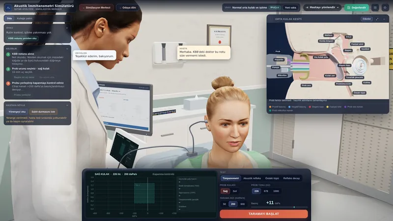
- Tympanometry: the obstructive type typically gives a type C tympanogram with peak pressure shifted to negative values; if fluid has collected, the trace flattens (type B with normal ear canal volume). In the baro-challenge-induced type the tympanogram is usually normal between episodes (type A). In the patulous type, fluctuations in time with breathing may be seen.
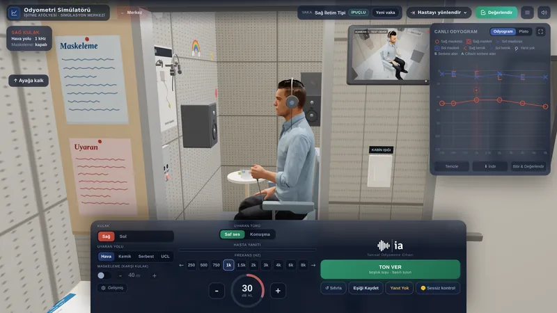
- Pure-tone audiometry: hearing may be normal, or there may be a mild conductive loss, mainly at low frequencies. Bone conduction is normal; any sensorineural component calls for a search for other causes.
- Acoustic reflexes: may be absent with marked negative pressure or effusion; they should be measured at the tympanometric peak pressure.
- ETDQ-7 questionnaire: the only patient-reported measure to have undergone validation (Schilder et al., 2015). It has seven items, each scored from 1 to 7, and in the original study a total score above 14.5 was taken as consistent with dysfunction (McCoul et al., 2012). In the Turkish adaptation, internal consistency (Cronbach’s alpha) was 0.714, and no healthy control scored above this threshold (Özgür et al., 2018).
- Eustachian tube function tests: tubomanometry, the nine-step inflation-deflation test and sonotubometry are used in research and specialist centres; there are no standardised diagnostic criteria yet.
- Nasopharyngoscopy and imaging: endoscopy may reveal an underlying cause. Imaging is not routine and is reserved for suspected additional pathology (Schilder et al., 2015).
| Feature | Obstructive | Baro-challenge-induced | Patulous |
|---|---|---|---|
| Problem | Tube fails to open | Cannot cope with pressure change | Tube stays open |
| When symptoms occur | Constant or fluctuating | Flying, diving | Exercise, after weight loss |
| Typical complaint | Fullness, muffled hearing | Pain, fullness | Autophony, hearing own breathing |
| Tympanometry | Negative pressure (C) or B | Usually normal (A) | Fluctuation with breathing |
| Diagnosis | Symptoms plus retraction or negative pressure | History | Symptoms plus drum movement with breathing |
The differential diagnosis includes other causes of aural fullness: earwax impaction, otitis media with effusion, patulous Eustachian tube, Ménière’s disease and sudden sensorineural hearing loss, superior semicircular canal dehiscence and temporomandibular joint problems. Measuring bone conduction thresholds is important so that inner ear causes of fullness are not missed.
Treatment and audiological rehabilitation
Acute dysfunction often resolves as an upper respiratory infection clears, and watchful waiting may be enough. Treating underlying nasal problems (allergic rhinitis, rhinosinusitis), stopping smoking and self-inflation (autoinflation) exercises are commonly advised; the use of nasal sprays or other medicines is decided by the doctor. A 2014 systematic review of interventions for adult Eustachian tube dysfunction concluded that the evidence at that time was insufficient to draw conclusions about the effectiveness of any intervention (Llewellyn et al., 2014).
Balloon dilation widens the tube by inflating a balloon placed through the nose into its cartilaginous part. Two randomised controlled trials form the core of the evidence. In a 60-patient trial, ETDQ-7 scores at six weeks fell by a mean of 2.9 points with balloon dilation and 0.6 points with medical management alone, and the benefit persisted to 12 months (Meyer et al., 2018). In a 323-patient trial, the tympanogram had normalised at six weeks in 51.8% after balloon dilation and in 13.9% of controls (Poe et al., 2018). In the UK, the National Institute for Health and Care Excellence (NICE) judged the evidence adequate to support the procedure with standard arrangements for clinical governance, consent and audit; the committee also noted that there was little evidence on the benefit of repeat procedures (NICE, 2019).
Persistent negative pressure, effusion or progressive retraction may be treated with a ventilation tube (grommet), which ventilates the middle ear independently of the tube but does not treat the underlying dysfunction and may extrude spontaneously. Grommets are also used selectively for frequent flyers or divers with baro-challenge-induced dysfunction; the decision rests with the surgeon. During follow-up the audiologist repeats tympanometry and audiometry to document change, and completing the ETDQ-7 before and after treatment gives an objective measure of benefit. Hearing aids are rarely needed.
Impact on quality of life and advice
Persistent fullness and muffled hearing can make meetings and phone calls harder, and constantly trying to clear the ears is tiring. Baro-challenge-induced dysfunction can make flying and diving painful, which matters for people who fly or dive for work. Knowing that the symptoms are not dangerous and understanding the follow-up plan helps to reduce anxiety.
When flying, swallow often, chew gum or sip water, especially once the descent begins; staying awake makes it easier to clear your ears. Babies can be fed from the breast or a bottle during descent. Avoid flying and diving with a blocked nose or a cold where possible, and ask your doctor if it cannot be avoided. When diving, equalise early and often from the surface and do not descend further if you feel pressure. Do not blow your nose forcefully; close one nostril and clear the other gently. Stopping smoking reduces mucosal swelling. Ask for reassessment if symptoms last more than three months or persist in one ear.
If you used this review, you can cite it as follows (APA 7):
Auditory Scene. (2026). Eustachian Tube Dysfunction. Hearing & Balance Health Guide. https://auditoryscene.com/guncel-haberler/categories/isitme-sagligi-rehberi/ostaki-borusu-disfonksiyonu/Permanent link: auditoryscene.com/guncel-haberler/categories/isitme-sagligi-rehberi/ostaki-borusu-disfonksiyonu/ · Last reviewed: October 2026 · License: CC BY-NC-ND 4.0
References
- Llewellyn, A., Norman, G., Harden, M., et al. (2014). Interventions for adult Eustachian tube dysfunction: A systematic review. Health Technology Assessment, 18(46).
- McCoul, E. D., Anand, V. K., & Christos, P. J. (2012). Validating the clinical assessment of eustachian tube dysfunction: The Eustachian Tube Dysfunction Questionnaire (ETDQ-7). The Laryngoscope, 122(5), 1137-1141.
- Meyer, T. A., O’Malley, E. M., Schlosser, R. J., et al. (2018). A randomized controlled trial of balloon dilation as a treatment for persistent eustachian tube dysfunction with 1-year follow-up. Otology & Neurotology, 39(7), 894-902.
- National Institute for Health and Care Excellence. (2019). Balloon dilation for chronic eustachian tube dysfunction (Interventional procedures guidance IPG665). NICE.
- Özgür, E., Bilgen, C., & Cengiz Özyurt, B. (2018). Turkish validity and reliability of Eustachian tube dysfunction questionnaire-7. Brazilian Journal of Otorhinolaryngology, 84(4), 435-440.
- Poe, D., Anand, V. K., Dean, M., et al. (2018). Balloon dilation of the eustachian tube for dilatory dysfunction: A randomized controlled trial. The Laryngoscope, 128(5), 1200-1206.
- Schilder, A. G. M., Bhutta, M. F., Butler, C. C., et al. (2015). Eustachian tube dysfunction: Consensus statement on definition, types, clinical presentation and diagnosis. Clinical Otolaryngology, 40(5), 407-411.
- Shan, A., Ward, B. K., Goman, A. M., et al. (2019). Prevalence of eustachian tube dysfunction in adults in the United States. JAMA Otolaryngology-Head & Neck Surgery, 145(10), 974-975.
Frequently asked questions
Does Eustachian tube dysfunction go away on its own?
Often, yes. Symptoms that start after a cold or an allergy flare usually ease within a few weeks. If they last more than three months, the dysfunction is considered chronic and an ear, nose and throat assessment is advisable.
Is it harmful to keep doing the Valsalva manoeuvre to clear my ears?
Gentle autoinflation is generally safe and may be recommended. Blowing very hard with the nose and mouth closed, however, can damage the eardrum and inner ear. Learn the technique from your doctor or audiologist, and stop and seek advice if you get pain, dizziness or reduced hearing.
What is the ETDQ-7 and why am I asked to complete it?
The ETDQ-7 scores Eustachian tube symptoms with seven questions, and a Turkish version has been validated (Özgür et al., 2018). It does not make the diagnosis on its own, but it helps to measure how severe your symptoms are and how they change after treatment.
Is balloon dilation a permanent solution?
In randomised trials, balloon dilation improved symptoms more than medical treatment alone, and the benefit lasted at least a year (Meyer et al., 2018). However, long-term evidence and evidence on repeat procedures are limited, and it is not suitable for everyone. The choice is made with your surgeon based on examination and tests.
What should I do before a flight?
If your nose is blocked or you have a cold, consider postponing the trip. During the flight, especially on descent, swallow often, chew gum and stay awake. For people who fly often and have severe pain on every flight, the doctor may suggest additional measures; any medicines are a decision for your doctor.
Can this affect my hearing permanently?
Hearing loss from Eustachian tube dysfunction is usually mild and temporary. However, long-standing negative pressure can cause permanent retraction pockets and, rarely, cholesteatoma. This is why regular examination and audiological follow-up matter when symptoms persist.
A questionnaire with a validated Turkish version that can be used to track the severity of Eustachian tube symptoms and the response to treatment: