Auditory Scene
At a glance
Pulsatile tinnitus is tinnitus synchronous with the heartbeat; it usually arises from turbulent blood flow in the arteries or veins around the temporal bone.
A beating or whooshing sound in time with the pulse is heard in one ear or bilaterally; some patients also have headache, visual symptoms or hearing loss.
Sudden onset, neurological signs, headache with visual disturbance or a bruit on examination need prompt assessment; some causes carry a risk of brain haemorrhage.
Most patients are imaged, and treatment depends on the cause: weight loss and medication, endovascular treatment, surgery, or observation with tinnitus counselling.
Medical disclaimer. This content is for informational purposes only and does not replace a physician’s examination, diagnosis or treatment; it should not be used as medical advice. For your complaints or personal situation, always consult an ear, nose and throat physician and an audiologist.
If you hear a sound in time with your heartbeat, see an ear, nose and throat doctor and say so clearly. Go to the emergency department without delay if the sound started suddenly, if you have recently had a head or neck injury, or if there is severe or worsening headache, blurred or double vision, slurred speech or weakness of an arm or leg. These symptoms may point to conditions affecting the blood vessels of the brain or the pressure inside the skull that need prompt treatment.
Definition and epidemiology
Pulsatile tinnitus is tinnitus that is synchronous with the heartbeat. Patients describe it as beating, whooshing or rushing in the ear, or as ‘hearing my heart in my ear’. If only the patient can hear it, it is subjective pulsatile tinnitus; if the examiner can hear it too, it is objective. Objective tinnitus is rare and may be caused by identifiable conditions such as vascular abnormalities and myoclonus (Tunkel et al., 2014). Rhythmic clicking that is not in time with the heartbeat suggests a muscular cause such as middle ear myoclonus instead.
Tinnitus affects 10-15% of adults, and pulsatile tinnitus makes up a small part of this group. The American tinnitus guideline excludes pulsatile tinnitus from its scope and, while recommending against routine imaging in tinnitus, lists pulsatile tinnitus, tinnitus localised to one ear, focal neurological abnormalities and asymmetric hearing loss as exceptions (Tunkel et al., 2014). Imaging reveals a finding in 57-100% of patients with pulsatile tinnitus, although the yield falls by 20-30% when debatable findings such as jugular bulb variants are excluded (Vattoth et al., 2010). For the general features of tinnitus, see tinnitus.
Symptoms and signs
The sound is usually heard in one ear and sometimes bilaterally. It may become more noticeable in quiet surroundings, when lying down or after exertion. Pulsatile tinnitus of venous origin is often a whooshing or rushing sound that may lessen with light pressure on the same side of the neck or with turning the head, whereas an arterial sound is sharper and more like a beat. A venous hum is also commonly heard in people without symptoms (Vattoth et al., 2010).
Associated symptoms give important clues to the cause. Headache, transient visual obscurations, double vision and blurred vision suggest idiopathic intracranial hypertension (IIH) (Mollan et al., 2018). Aural fullness, hearing loss and a red mass behind the eardrum suggest glomus tumours (paragangliomas); a blue mass behind the drum may indicate a dehiscent jugular bulb, and a red mass a persistent stapedial artery (Vattoth et al., 2010); an aberrant internal carotid artery can also appear as a mass behind the drum. Hearing one’s own voice and eye movements, sound-induced dizziness and a low-frequency air-bone gap suggest superior semicircular canal dehiscence. Sudden onset, neck pain or neurological symptoms are warning signs of carotid dissection or a dural arteriovenous fistula.
Causes and risk factors
Causes fall into two broad groups, arterial and venous. Arterial causes include carotid stenosis and dissection, fibromuscular dysplasia, an aberrant or dehiscent internal carotid artery, a persistent stapedial artery and arteriovenous malformations; highly vascular paragangliomas are considered in this group as well (Vattoth et al., 2010). Dural arteriovenous fistulas (DAVF) are the commonest cause of objective pulsatile tinnitus in patients with normal otoscopy; they usually lie in the transverse, sigmoid and cavernous sinuses and are supplied by branches of the external carotid artery (Vattoth et al., 2010). Their significance lies in the pattern of venous drainage: type I fistulas with antegrade drainage into a sinus follow a benign course, whereas reflux into cortical veins or direct cortical drainage markedly increases the risk of haemorrhage, reported in 10% of type IIb, 40% of type III and 65% of type IV fistulas (Cognard et al., 1995).
Venous causes include a dominant jugular system, a high or dehiscent jugular bulb, sigmoid sinus diverticulum and dehiscence, transverse sinus stenosis and IIH. Sigmoid sinus wall anomalies are a recognised cause of pulsatile tinnitus and can be repaired surgically (Eisenman et al., 2018). IIH occurs mainly in women with obesity, and pulsatile tinnitus is among its possible symptoms together with headache and visual complaints (Mollan et al., 2018). In patients with IIH, severe transverse sinus stenosis was found in 49% and sigmoid sinus dehiscence in 41% (compared with 7% and 27% of controls), but these findings did not differ between IIH patients with and without pulsatile tinnitus and may not be its direct structural cause (Lansley et al., 2017). Otosclerosis and Paget’s disease can also cause pulsatile tinnitus (Vattoth et al., 2010), and high-output states such as anaemia and hyperthyroidism should be borne in mind.
Audiological and clinical assessment
Assessment begins with a detailed history: synchrony with the heartbeat is confirmed by feeling the pulse, and the side, mode of onset, change with neck pressure or head position, headache, visual symptoms, trauma, weight gain and cardiovascular disease are explored. The guideline recommends a thorough medical evaluation in pulsatile tinnitus to exclude systemic cardiovascular or neurological disease (Tunkel et al., 2014).
- Otoscopy: look for a red or blue mass behind the drum; a vascular mass should not be touched or pressurised.
- Auscultation: listen over the ear, mastoid, orbit and neck with a stethoscope. A bruit that the examiner can hear indicates objective pulsatile tinnitus and raises the possibility of causes such as a dural arteriovenous fistula.
- Neck pressure and head turning: a reduction in the sound with light pressure on the ipsilateral jugular vein or with head turning suggests a venous source.
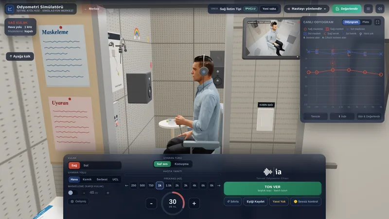
- Pure-tone audiometry: usually normal. A conductive loss suggests a middle ear mass or otosclerosis; a low-frequency air-bone gap with normal or better-than-normal bone conduction suggests superior semicircular canal dehiscence; and a sensorineural loss points to inner ear or internal auditory canal pathology.
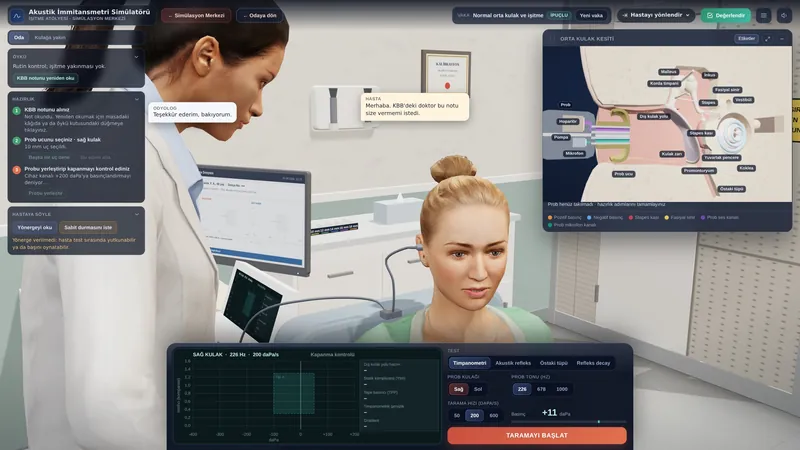
- Tympanometry and acoustic reflexes: help to separate middle ear masses, otosclerosis and dehiscence; fluctuations synchronous with the heartbeat during recording support a vascular source.
- Other tests: vestibular evoked myogenic potentials (VEMP) when dehiscence is suspected; fundoscopy and visual fields when IIH is suspected.
| Finding | Causes to consider | First investigation |
|---|---|---|
| Red mass behind the drum | Paraganglioma, aberrant carotid | Contrast temporal bone CT and MRI |
| Normal otoscopy, venous features | Sigmoid sinus anomaly, sinus stenosis | CT angiography and venography |
| Objective bruit, neurological signs | Dural AV fistula, AVM | MR angiography, DSA |
| Headache, visual symptoms | IIH, venous sinus thrombosis | MRI and MRV, eye examination |
| Neck pain, sudden onset | Carotid dissection | Neck CTA or MRA |
There is general consensus that contrast-enhanced high-resolution CT of the temporal bone is the first imaging study. If CT is normal, further investigation reveals a finding in 30-40% of patients: carotid ultrasound for stenosis, neck CT angiography (CTA) for dissection and masses, and conventional angiography for dural fistulas. Time-resolved MR angiography can show most vascular lesions, and digital subtraction angiography (DSA) is the technique of choice when an arteriovenous malformation or fistula is suspected (Vattoth et al., 2010). CT arteriography and venography can assess arterial, venous, middle ear and inner ear causes in a single examination (Krishnan et al., 2006). When IIH is suspected, MRI and CT or MR venography (MRV) to exclude venous sinus thrombosis should be performed within 24 hours, followed, if imaging is normal, by lumbar puncture to measure the opening pressure; values above 25 cm CSF support the diagnosis (Mollan et al., 2018).
Treatment and audiological rehabilitation
Treatment depends on the cause, and decisions are made together with the relevant specialists. In IIH, weight loss is the only disease-modifying treatment, and medication may be used for symptoms. Fulminant IIH with rapidly declining vision needs urgent surgery to preserve sight (Mollan et al., 2018). Dural arteriovenous fistulas with cortical venous drainage should be treated because of the risk of haemorrhage; classifying the venous drainage helps to estimate the risk of each fistula and guide treatment (Cognard et al., 1995). These fistulas are usually treated endovascularly. For glomus tumours, observation, surgery or radiotherapy are considered.
Pulsatile tinnitus due to sigmoid sinus wall anomalies can be treated by repairing the sinus wall through a transmastoid approach (Eisenman et al., 2018). Carotid stenosis and dissection are managed by vascular surgery and neurology, and treating systemic causes such as anaemia and hyperthyroidism can also relieve pulsatile tinnitus. In superior semicircular canal dehiscence, surgical repair is considered when symptoms are severe.
When imaging shows no cause that needs treatment, the approach resembles tinnitus management: explaining the source of the sound and that it is harmless, tinnitus counselling, sound therapy and hearing aids if there is an accompanying hearing loss. At this stage the audiologist documents tinnitus severity with questionnaires and, if anything changes during follow-up, such as a change in the character of the sound or a new neurological symptom, refers the patient back for reassessment.
Impact on quality of life and advice
Because pulsatile tinnitus is most noticeable at night, in quiet rooms and when lying down, it can make it hard to fall asleep. Hearing one’s heartbeat constantly can cause anxiety, and some patients fear they have a serious illness. Investigating the cause and explaining the results clearly reduces this anxiety considerably. In conditions such as IIH, headache and visual problems also affect quality of life.
Practical advice: note when the sound gets louder or quieter and whether it changes with neck pressure or head position, and tell your doctor. Background sound at night, such as a fan, soft music or nature sounds, makes the sound less intrusive. If you have been diagnosed with IIH, joining a weight management programme and keeping your eye appointments are important. Seek help without waiting if the character of the sound changes or if new headache, visual or speech problems appear.
If you used this review, you can cite it as follows (APA 7):
Auditory Scene. (2026). Pulsatile Tinnitus. Hearing & Balance Health Guide. https://auditoryscene.com/guncel-haberler/categories/isitme-sagligi-rehberi/pulsatil-tinnitus/Permanent link: auditoryscene.com/guncel-haberler/categories/isitme-sagligi-rehberi/pulsatil-tinnitus/ · Last reviewed: October 2026 · License: CC BY-NC-ND 4.0
References
- Cognard, C., Gobin, Y. P., Pierot, L., et al. (1995). Cerebral dural arteriovenous fistulas: Clinical and angiographic correlation with a revised classification of venous drainage. Radiology, 194(3), 671-680.
- Eisenman, D. J., Raghavan, P., Hertzano, R., et al. (2018). Evaluation and treatment of pulsatile tinnitus associated with sigmoid sinus wall anomalies. The Laryngoscope, 128(S2), S1-S13.
- Krishnan, A., Mattox, D. E., Fountain, A. J., et al. (2006). CT arteriography and venography in pulsatile tinnitus: Preliminary results. American Journal of Neuroradiology, 27(8), 1635-1638.
- Lansley, J. A., Tucker, W., Eriksen, M. R., et al. (2017). Sigmoid sinus diverticulum, dehiscence, and venous sinus stenosis: Potential causes of pulsatile tinnitus in patients with idiopathic intracranial hypertension? American Journal of Neuroradiology, 38(9), 1783-1788.
- Mollan, S. P., Davies, B., Silver, N. C., et al. (2018). Idiopathic intracranial hypertension: Consensus guidelines on management. Journal of Neurology, Neurosurgery & Psychiatry, 89(10), 1088-1100.
- Tunkel, D. E., Bauer, C. A., Sun, G. H., et al. (2014). Clinical practice guideline: Tinnitus. Otolaryngology-Head and Neck Surgery, 151(2_suppl), S1-S40.
- Vattoth, S., Shah, R., & Curé, J. K. (2010). A compartment-based approach for the imaging evaluation of tinnitus. American Journal of Neuroradiology, 31(2), 211-218.
Frequently asked questions
I can hear my heartbeat in my ear. Is it dangerous?
It usually has a harmless cause, but unlike other types of tinnitus it is sometimes due to a blood vessel or pressure problem that needs treatment. That is why assessment by an ENT doctor is recommended and, in most cases, imaging.
Which tests will I have?
Ear examination, listening over the ear and neck with a stethoscope, a hearing test and tympanometry come first. CT, MRI or vascular imaging follows depending on the findings. If you have headaches and visual symptoms, an eye examination is also needed.
The sound gets quieter when I press on my neck. What does that mean?
This suggests that the sound comes from blood flow in the veins. Thinning or a gap in the wall of the sigmoid sinus, narrowing of a venous sinus and raised pressure inside the skull belong to this group. Make sure you tell your doctor about this observation.
What is idiopathic intracranial hypertension?
It is a condition in which the pressure of the cerebrospinal fluid rises without an obvious cause; it mostly affects women with obesity. It can cause headaches, visual problems and pulsatile tinnitus. Because it can threaten sight, eye checks are important, and weight loss is the basis of treatment.
My scans showed no cause. What can I do?
The approach is then similar to tinnitus management: knowing that the sound is harmless, counselling, using background sound at night and sound therapy if needed ease the problem. If the character of the sound changes or new symptoms appear, ask to be seen again.
Does pulsatile tinnitus cause hearing loss?
The sound itself does not harm hearing. Some causes, however, such as a glomus tumour, otosclerosis or semicircular canal dehiscence, can also cause hearing loss, so a hearing test is a core part of the assessment.
A scale that can be used to assess and monitor the impact of pulsatile tinnitus on daily life: