Auditory Scene
At a glance
A temporal bone fracture is a break in the skull base bone that houses the ear and facial nerve; today fractures are classified as otic capsule sparing or otic capsule violating.
Bleeding from the ear, hearing loss, dizziness, facial weakness and clear fluid from the ear or nose are the main signs.
It is an emergency: initial care targets life-threatening injuries, and facial movement should be documented in the emergency department.
CSF leaks and facial palsy are observed or treated surgically by doctors; hearing is monitored audiologically, and persistent loss is managed with surgery, hearing aids or implants.
Medical disclaimer. This content is for informational purposes only and does not replace a physician’s examination, diagnosis or treatment; it should not be used as medical advice. For your complaints or personal situation, always consult an ear, nose and throat physician and an audiologist.
After a head injury, go to the emergency department straight away and call 112 if there is bleeding from the ear, clear fluid from the ear or nose, weakness of one side of the face, sudden hearing loss or severe dizziness. Do not try to clean, syringe or put drops into the ear. After discharge, keep your ENT and audiology appointments if your hearing has not recovered or if tinnitus or unsteadiness persists.
Definition and epidemiology
The temporal bone contains the ear canal, middle ear, inner ear (otic capsule), facial nerve, internal carotid artery and sigmoid sinus. Breaking it takes considerable force: cadaver studies show that a lateral impact of 6,000-8,000 N is needed. In the largest series, 31% of fractures were caused by road traffic collisions, followed by assaults and falls; men are affected 3-4 times as often as women, fractures are bilateral in 8-29% of patients and about 60% are open (Diaz et al., 2016). A UK major trauma centre identified 815 cases in about six and a half years and emphasised that these fractures indicate high-energy trauma (Kanona et al., 2020).
The traditional classification describes the orientation of the fracture line relative to the long axis of the petrous pyramid: longitudinal fractures account for 70-90% and transverse fractures for 10-30% (Kurihara et al., 2020). Most fractures have mixed features, however, and this distinction predicts complications poorly (Diaz et al., 2016; Ishman and Friedland, 2004). The classification proposed by Dahiya and colleagues separates otic capsule sparing from otic capsule violating fractures; the latter make up 5-20% of all fractures (Dahiya et al., 1999; Kurihara et al., 2020). Distinguishing petrous from non-petrous fractures likewise correlates better with clinical outcomes (Ishman and Friedland, 2004).
Symptoms and signs
In the acute phase there may be bleeding from the ear, a laceration or step in the ear canal, haemotympanum (blood behind the eardrum), hearing loss, tinnitus, dizziness and nystagmus. Bruising over the mastoid (Battle’s sign) and around the eyes suggests a skull base fracture. Clear fluid from the ear or nose may indicate a cerebrospinal fluid (CSF) leak, which occurs in 17% of fractures (Diaz et al., 2016). Because of reduced consciousness and associated brain injury, ear findings can be missed in the first days.
Facial palsy develops in 6-7% of temporal bone fractures and in 3-9% in children. In 27% of cases the palsy is present from the moment of injury; in 73% facial movement is initially preserved and then deteriorates within 1-16 days (Diaz et al., 2016). In a series of 820 fractures there were 58 facial nerve injuries; all incomplete palsies and all but one of the delayed-onset palsies recovered well, whereas 40% of immediate complete palsies had a poor recovery (Brodie and Thompson, 1997). Otic capsule violating fractures are more than twice as likely to cause facial palsy, four times as likely to cause a CSF leak and seven times as likely to cause profound hearing loss (Dahiya et al., 1999; Kurihara et al., 2020).
Causes and risk factors
The direction of the fracture depends on the site and force of the impact. Lateral blows usually produce otic capsule sparing fractures through the ear canal and middle ear, in which blood and ossicular injury cause a conductive or mixed loss. Ossicular chain injury is found in about 20% of cases; the commonest injuries are incudostapedial joint separation (82%), incus dislocation (57%) and fracture of the stapes crura (30%) (Diaz et al., 2016). Ossicular dislocations are more common than fractures, and among fractures the long process of the incus is most often affected (Kurihara et al., 2020). These injuries are described in detail under ossicular chain discontinuity.
Otic capsule violating fractures cut directly through the cochlea and labyrinth and almost always cause sensorineural hearing loss and vertigo (Diaz et al., 2016). Even without a fracture of the capsule, the impact can concuss the membranous labyrinth; labyrinthine concussion presents with sudden sensorineural loss and vertigo, and inner ear haemorrhage may be visible on MRI (Kurihara et al., 2020). Half of ears with traumatic incus dislocation also have a sensorineural component of 10 dB or more (Diaz et al., 2016). Injury to the oval or round window can cause a perilymph fistula, and displaced otoconia can cause post-traumatic positional vertigo. The facial nerve is most often injured around the geniculate ganglion. Tegmen defects predispose to CSF leaks, and fractures crossing the carotid canal to internal carotid artery injury (Kurihara et al., 2020).
Audiological and clinical assessment
Until the patient is stable, assessment focuses on life-threatening injuries. Documenting facial movement in the emergency department is critical for distinguishing immediate from delayed palsy (Brodie and Thompson, 1997). In the acute phase hearing is assessed at the bedside with whispered voice and tuning forks; pure-tone and speech audiometry are usually deferred unless surgery is planned (Diaz et al., 2016).
- Otoscopy: look for canal laceration or step, haemotympanum, perforation and otorrhoea (ear discharge). Clear discharge is assessed for CSF; the ear is not syringed and no instruments are inserted.
- Tuning fork tests: a Weber that lateralises to the injured ear suggests a conductive loss, and one that lateralises to the other ear suggests a sensorineural loss.
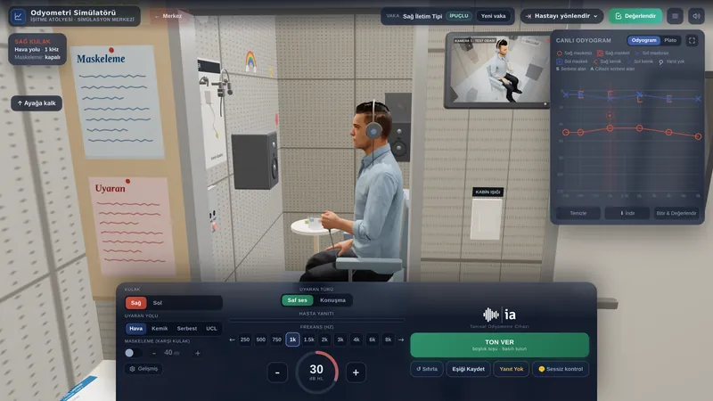
- Pure-tone audiometry: haemotympanum produces a conductive loss with an air-bone gap that resolves over days to weeks as the blood clears. A high-frequency drop in bone conduction points to labyrinthine concussion or otic capsule injury; with an otic capsule violating fracture the loss is usually severe or profound.
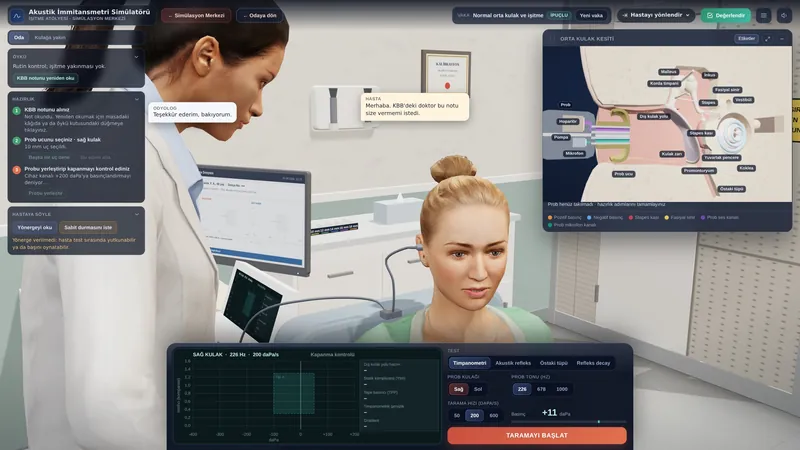
- Tympanometry and acoustic reflexes: haemotympanum gives a flat (type B) trace and ossicular discontinuity a high-compliance (type Ad) trace; a perforation gives a high volume. If the middle ear is normal, an absent stapedial reflex on the side of a facial palsy suggests a lesion proximal to the nerve to stapedius.
- Facial nerve tests: in immediate complete palsy, electroneurography (ENoG) is performed on days 3-7; degeneration above 90% within 6 days or above 95% within 14 days supports surgical exploration (Diaz et al., 2016).
- Vestibular tests: the Dix-Hallpike and supine roll tests identify post-traumatic BPPV, while the video head impulse test (vHIT), caloric testing and vestibular evoked myogenic potentials (VEMP) show labyrinthine damage.
Thin-section CT (1 mm or less) with a bone algorithm is the core investigation; CT or MR angiography is recommended when the fracture crosses the carotid canal. Air in the inner ear (pneumolabyrinth) strongly suggests an otic capsule fracture or a perilymph fistula, and MRI can show inner ear haemorrhage in unexplained sensorineural loss or vertigo (Kurihara et al., 2020). Whereas the traditional classification did not significantly predict complications, the otic capsule system did: otic capsule violating fractures were 5 times more likely to cause facial nerve injury, 25 times more likely to cause sensorineural loss and 8 times more likely to cause CSF otorrhoea (Little and Kesser, 2006).
| Feature | Otic capsule sparing | Otic capsule violating |
|---|---|---|
| Frequency | Most fractures | 5-20% |
| Hearing loss | Conductive or mixed | Sensorineural, usually severe |
| Facial palsy | Less common | 2-5 times more common |
| CSF leak | Less common | 4-8 times more common |
| Vertigo | Mostly BPPV, transient | From labyrinthine damage, marked |
Treatment and audiological rehabilitation
Management is multidisciplinary and decisions rest with the doctors. Between 57% and 85% of CSF leaks stop within a week with conservative care, and surgical repair is recommended for leaks lasting more than 7-10 days (Diaz et al., 2016). In a series of 122 CSF fistulas, 95 closed spontaneously within a week; the risk of meningitis rose to 23% in fistulas lasting more than seven days, compared with 3% in those that closed earlier (Brodie and Thompson, 1997). Delayed-onset facial palsy usually recovers with steroids and observation; in immediate complete palsy, surgical decompression is discussed according to electrophysiological findings (Diaz et al., 2016). High-dose steroids are not advised in patients with intracranial injury (Kanona et al., 2020).
Conductive loss due to haemotympanum usually resolves on its own. If a 30 dB loss persists beyond two months, ossicular injury is suspected and exploration with ossiculoplasty is considered; the air-bone gap closes to within 10 dB in 78% of cases. Surgery is not performed on an only hearing ear (Diaz et al., 2016); in that situation, or when surgery is not wanted, a hearing aid is a good option. For profound sensorineural loss after an otic capsule violating fracture, CROS, bone conduction implants and, in suitable cases, a cochlear implant (CI) are considered; before cochlear implantation, cochlear patency and the cochlear nerve are assessed by imaging.
Most post-traumatic vertigo settles within 4-6 weeks; the commonest cause is BPPV, which is treated with repositioning manoeuvres. When a perilymph fistula is suspected, observation for at least 1-2 weeks is recommended (Diaz et al., 2016). Persistent unilateral vestibular loss is managed with vestibular rehabilitation. Hearing and balance problems after head injury without a fracture are covered under hearing and balance problems after head injury.
Impact on quality of life and advice
A temporal bone fracture is usually part of a major injury, and hearing and balance problems can be overshadowed by brain injury, orthopaedic injuries and a long hospital stay. Planned audiological follow-up after discharge is therefore important: the first full assessment is made once the patient can take part in testing, and repeated audiograms show whether the conductive loss is resolving and whether any sensorineural component is progressing. A unilateral loss makes it harder to follow speech in noise and to locate sounds, which can affect the return to work or school.
Facial palsy affects eye closure, speech and eating and can cause concern about appearance; eye protection and physiotherapy are planned on medical advice. Dizziness and unsteadiness increase the risk of falls, and returning to driving or working at heights should be discussed with the doctor. Practical advice: seek help without delay if clear fluid starts to come from your ear, or if you develop fever, neck stiffness or severe headache; avoid blowing your nose and straining until your doctor says otherwise; keep your hearing and balance appointments and tell your audiologist about the difficulties you notice.
If you used this review, you can cite it as follows (APA 7):
Auditory Scene. (2026). Temporal Bone Fractures. Hearing & Balance Health Guide. https://auditoryscene.com/guncel-haberler/categories/isitme-sagligi-rehberi/temporal-kemik-kiriklari/Permanent link: auditoryscene.com/guncel-haberler/categories/isitme-sagligi-rehberi/temporal-kemik-kiriklari/ · Last reviewed: October 2026 · License: CC BY-NC-ND 4.0
References
- Brodie, H. A., & Thompson, T. C. (1997). Management of complications from 820 temporal bone fractures. The American Journal of Otology, 18(2), 188-197.
- Dahiya, R., Keller, J. D., Litofsky, N. S., et al. (1999). Temporal bone fractures: Otic capsule sparing versus otic capsule violating clinical and radiographic considerations. The Journal of Trauma, 47(6), 1079-1083.
- Diaz, R. C., Cervenka, B., & Brodie, H. A. (2016). Treatment of temporal bone fractures. Journal of Neurological Surgery Part B: Skull Base, 77(5), 419-429.
- Ishman, S. L., & Friedland, D. R. (2004). Temporal bone fractures: Traditional classification and clinical relevance. The Laryngoscope, 114(10), 1734-1741.
- Kanona, H., Anderson, C., Lambert, A., et al. (2020). A large case series of temporal bone fractures at a UK major trauma centre with an evidence-based management protocol. The Journal of Laryngology & Otology, 134(3), 205-212.
- Kurihara, Y. Y., Fujikawa, A., Tachizawa, N., et al. (2020). Temporal bone trauma: Typical CT and MRI appearances and important points for evaluation. RadioGraphics, 40(4), 1148-1162.
- Little, S. C., & Kesser, B. W. (2006). Radiographic classification of temporal bone fractures: Clinical predictability using a new system. Archives of Otolaryngology-Head & Neck Surgery, 132(12), 1300-1304.
Frequently asked questions
There is blood coming from my ear. Is my hearing permanently affected?
Not necessarily. Most of the blood comes from a tear in the ear canal or from blood collecting behind the eardrum, and the resulting conductive loss usually improves as the blood clears. When the fracture involves the inner ear, however, the hearing loss is usually permanent. Hearing tests and a CT scan show which applies.
Clear, watery fluid is coming from my ear. What should I do?
This may be cerebrospinal fluid and carries a risk of meningitis. Do not plug, clean or put drops into the ear; contact your doctor or go to the emergency department straight away. Most leaks stop on their own within a week; those that persist are repaired surgically.
Will my facial palsy recover?
If your face moved for a while after the injury and weakened later, or if the palsy is incomplete, the chance of recovery is very high. If the face was completely paralysed from the moment of injury, recovery may be more limited; nerve tests are then carried out and your doctor decides whether surgery is needed.
When should my hearing be tested?
Bedside tests are used while you are in intensive care; a full hearing test is done once you are able to take part. Tests are repeated until the blood behind the eardrum has cleared. If a conductive loss lasts longer than two months, the ENT doctor will reassess you for ossicular injury.
Since the accident the room spins when I turn my head. Is this due to the fracture?
BPPV is the commonest cause of dizziness after a head injury and can occur with or without a fracture. Brief dizziness triggered by head movement can be treated with simple manoeuvres; tell your doctor and audiologist about your symptoms.
What can be done if my hearing loss is permanent?
A conductive loss can be treated with ossicular reconstruction or a hearing aid, and an inner ear loss with a hearing aid. If one ear is left with profound hearing loss, a CROS aid, bone conduction implants or, where suitable, a cochlear implant can be considered.
Scales that can be used to follow the impact of residual hearing loss and dizziness on daily life after a fracture: